
Explaining the Different Types of Scans
BHD is characterised by 3 main manifestations: skin bumps called fibrofolliculomas, lung cysts and collapsed lungs and kidney cancer. Currently, there is no cure. Therefore, management of your symptoms through regular scans is important to minimise the risk of kidney cancer. However, you may also have scans for your lungs to check for lung cysts or if your lung collapses (called a pneumothorax).
In this toolkit, we will explain the different types of scans you may have and when and why you would have a particular type of scan. The information in this toolkit is meant to provide a general overview of the different types of scans but is not a substitute for medical advice. Please discuss any concerns with your doctor.
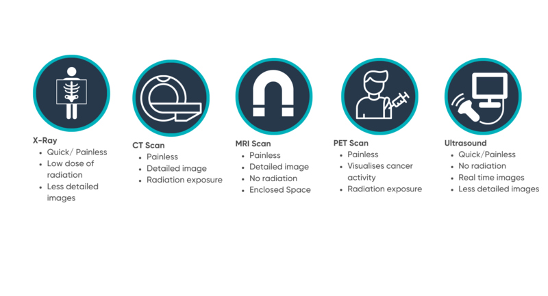
X-ray
These scans are quick and painless and use radiation to view structures within the body. The radiation passes through soft structures and is absorbed by harder structures (e.g. bones). The radiation passed through the body is measured by a detector and creates the final image. It is particularly good at visualising harder structures such as bone, but the images are not very detailed.
X-rays involve small doses of radiation equivalent to anywhere between a few days and few years of normal background radiation. During the procedure, the body part being scanned will be positioned against the radiation detector with the X-ray machine opposite. The scan will only take a couple of minutes and if done as an outpatient (i.e. not staying in hospital) you can usually go home immediately.
When would you have an X-ray?
You may have an X-ray of your chest to check if you have a collapsed lung (pneumothorax). This is the most common scan to diagnose a pneumothorax, Imaging such a CT scan (see below) may then be used to investigate further.
CT scan
Also known as a CAT scan, these scans involve multiple x-rays from different angles to create very detailed images of the inside of the body. They are used to diagnose, monitor, and guide treatment in several different conditions.
CT scans involve radiation equivalent to anywhere between a few days and few years of normal background radiation. During the scan you will be lying on a bed and moved through the scanner which looks like a giant donut (you will never be completely enclosed by the scanner and always be able to speak to someone). The scan normally lasts between 10-20 minutes depending on the part of the body being scanned. Sometimes, to get an even clearer picture of what is going on you will be given a dye (known as contrast). Depending on the reason for the scan the dye can be given as a drink, injected into a blood vessel or given as an enema (capsule put into the bottom). If you have a CT scan as an outpatient, you can usually go home immediately after but may be asked to wait for 30 minutes to an hour if you have had the dye as in rare cases (1 in 1000 people) it can cause an allergic reaction.
When would you have a CT scan?
Experts recommend that BHD patients should have a high-resolution CT scan when they are first diagnosed, to show how many cysts are present. There is no need for follow up scans if a patient has no further issues with their lungs. You may also have a CT scan if you have a collapsed lung or potentially to monitor your kidneys. However, getting CT scans regularly is not recommended due to the high doses of radiation.
MRI scan
An MRI scan is a safe painless procedure which uses magnetic fields and radio waves to create images of the body (no radiation). Like CT scans, the images produced are very detailed, however MRI scans are particularly good at visualising softer structures (e.g. the kidneys) in the body. The images can help to diagnose, monitor and manage a condition.
During an MRI scan you will be put on a bed and moved into the scanner either feet or head first depending on the scan. For an MRI scan of your abdomen, you normally enter the scanner feet first. The scanner is a long tube, some people do find them claustrophobic, but you will always be able to communicate with someone while inside. They also are very noisy, and you will be given earbuds or headphones to wear. In most cases, you can choose to listen to your own music. If you have concerns about this, always talk to your medical team as sometimes they can provide a light sedative. MRI scans range in length depending on the area being scanned and usually last between 20 minutes to an hour. Sometimes, to get an even clearer picture of what is going on you will be given a dye (known as contrast). The dye can be given as a drink, injected into a blood vessel or given as an enema (capsule put into the bottom). If you have an MRI scan as an outpatient, you can usually go home immediately after but may be asked to wait for 30 minutes to an hour if you have had the dye as in rare cases it can cause an allergic reaction.
When would you have an MRI scan?
If an MRI scan is available through your healthcare system, it is the recommended choice of scan to monitor your kidneys. How often you get these scans may vary depending on where you live and/or the size of any kidney tumours. In some countries you may get kidney scans yearly and in other it may be every 2 or 3 years. If you currently don’t have any kidney tumours, you may get scans less often than someone who has kidney tumours.
Ultrasound
An ultrasound is a quick, painless scan which uses sound waves to visualise structures in the body. It is the scan used on pregnant women to have a look at an unborn child. Although the images created from ultrasound are not very detailed, they are useful to visualise organs in real time and as it involves no radiation they are completely safe. They usually take between 15-25 minutes.
When would you have an ultrasound?
If an MRI scan to monitor your kidney isn’t available, you may have an ultrasound. However, ultrasounds aren’t sensitive enough to detect very small tumours.
PET Scan
A PET scan is a painless procedure used to visualise activity in your body rather than just creating images of the structures present. It is often used to investigate and monitor confirmed cases of cancer. The scan involves being injected with a substance which emits radiation (this is known as radiotracer). This substance acts similarly to glucose, the molecule that provides us with energy, and will build up in areas which use a lot of energy. Cancer cells need a lot of energy and therefore the radiotracer accumulates where the active cancer is and is interpreted by the PET scanner as an image. The resulting image shows up areas where the radiotracer is.
When would you have a PET scan?
It is unlikely you will ever have a PET scan, unless you have kidney cancer that has spread to other organs (metastasised). Kidney cancer associated with BHD is normally slow growing and rarely spreads. Early diagnosis and regular kidney scans will also allow any kidney cancer to be caught early.
Although the symptoms of BHD aren’t normally life threatening, management of the condition is critical to identify and treat any kidney cancer as early as possible. Currently, there are no standardised guidelines for the management of BHD. We know that it can be confusing as different people are told different things depending on your location and healthcare system and individual circumstances. Not only can the type of scan differ, but how often you get a scan can vary.
You can help advance our understanding of BHD
Find out howBack to